40 heart structure with labels
Easy way to draw heart structure by 5 steps | labeling of heart ... My youtube channel : facebook page : way to draw hea... Structure and function of the heart - BBC Bitesize The structure of the heart. If you clench your hand into a fist, this is approximately the same size as your heart. It is located in the middle of the chest and slightly towards the left.
Heart Diagram – 15+ Free Printable Word, Excel, EPS, PSD Teachers and students use the heart diagram, in biological science, to study the structure and functions of a human being’s heart. Friends and colleagues on the other hand may find this diagram template useful when it comes to sending special, personalized gifts to their family members and significant others. Download the template today, and ...

Heart structure with labels
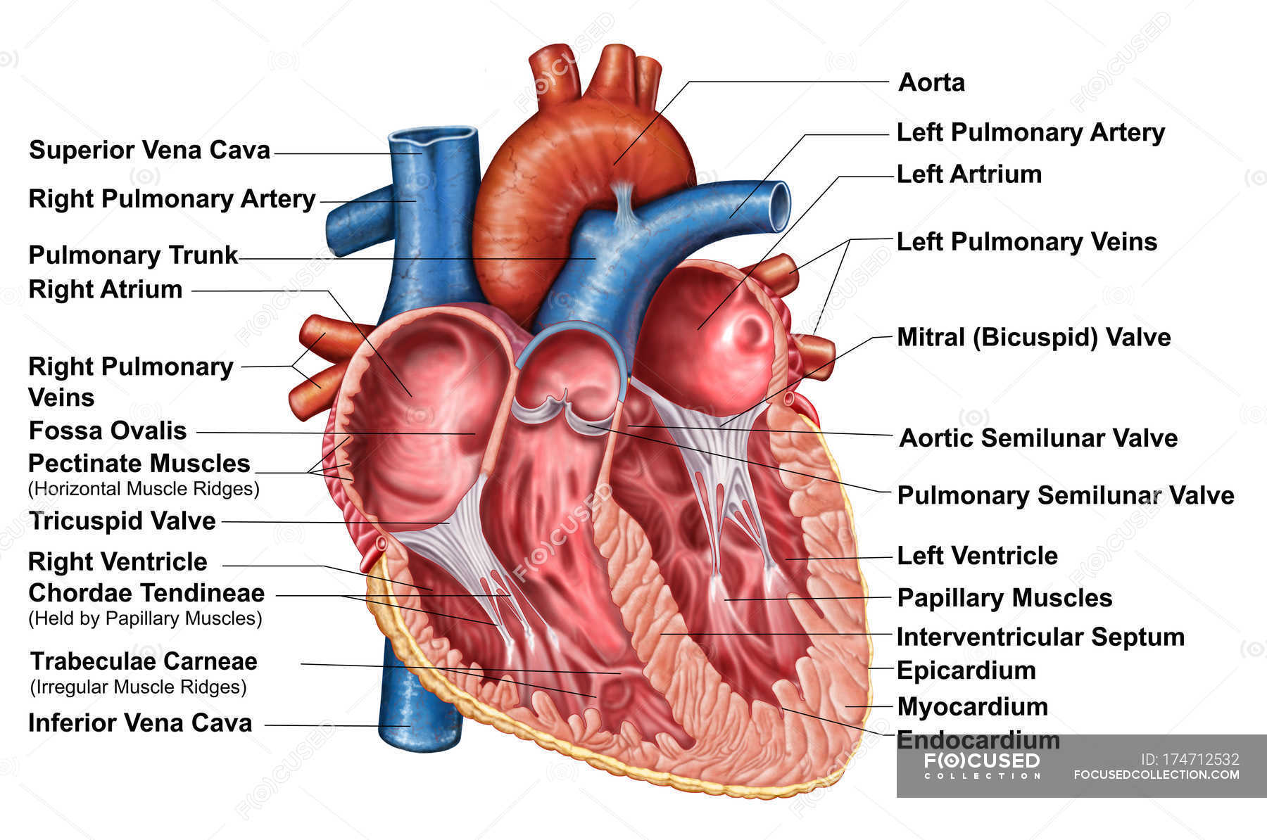
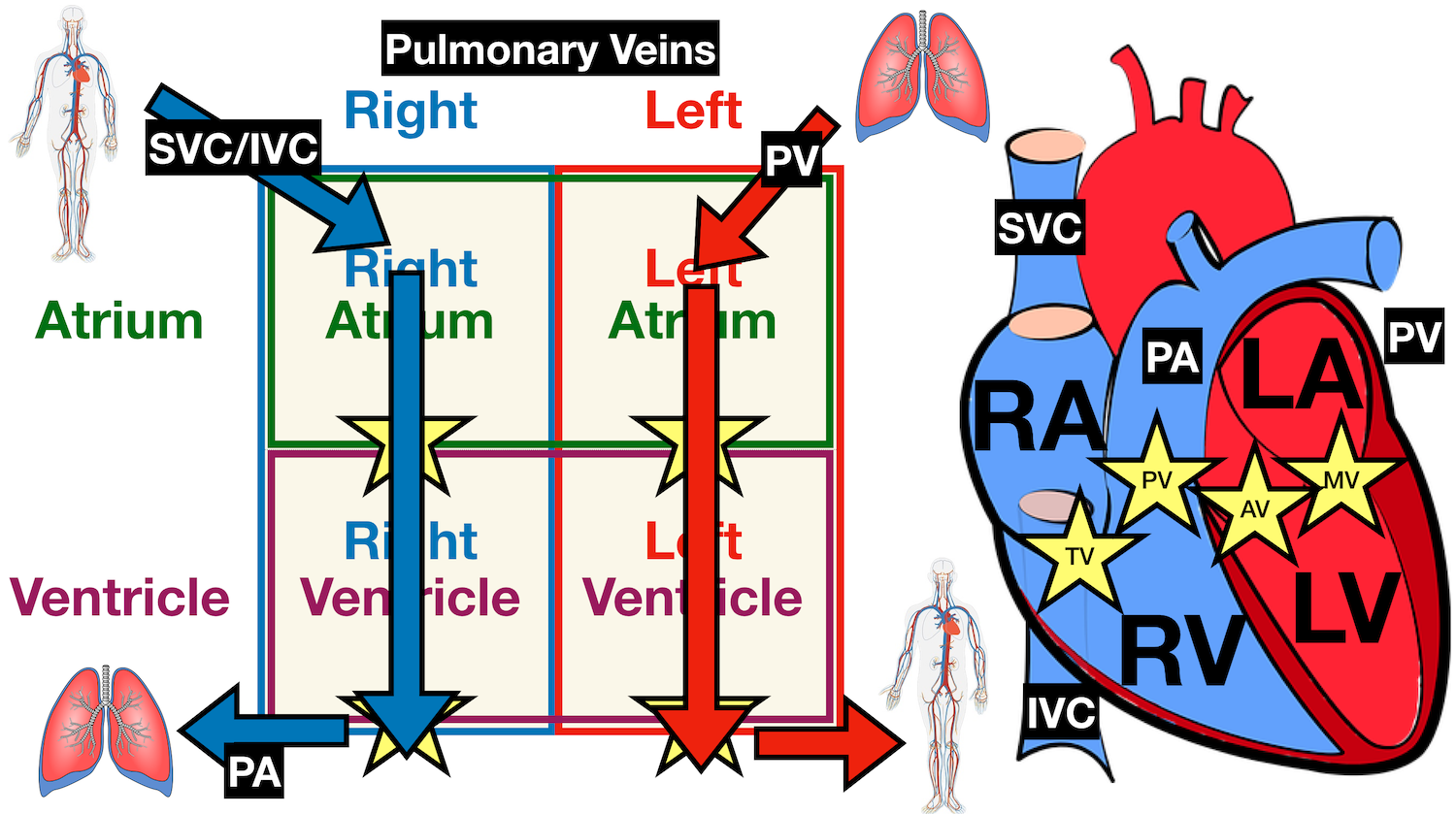
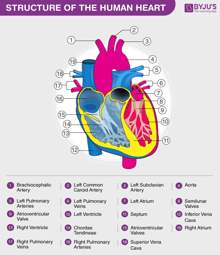
Heart Diagram with Labels and Detailed Explanation - BYJUS Diagram of Heart. The human heart is the most crucial organ of the human body. It pumps blood from the heart to different parts of the body and back to the heart. The most common heart attack symptoms or warning signs are chest pain, breathlessness, nausea, sweating etc. The diagram of heart is beneficial for Class 10 and 12 and is frequently ... Structure of the Heart | SEER Training - National Cancer Institute Layers of the Heart Wall Three layers of tissue form the heart wall. The outer layer of the heart wall is the epicardium, the middle layer is the myocardium, and the inner layer is the endocardium. Chambers of the Heart The internal cavity of the heart is divided into four chambers: Right atrium Right ventricle Left atrium Left ventricle RiSA planning overhaul of Samas 'structure, system and … 01.09.2022 · Sibisi attributed this to "less than optimal" engagement with their members (record companies and artist-led labels) because of Covid-19 and the government-imposed restrictions. "The Samas rely on a system in which our members' review, amend and confirm the rules, categories, and judges. This system is reviewed annually by our members and ...
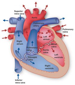
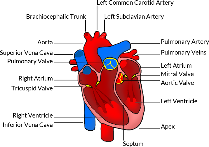
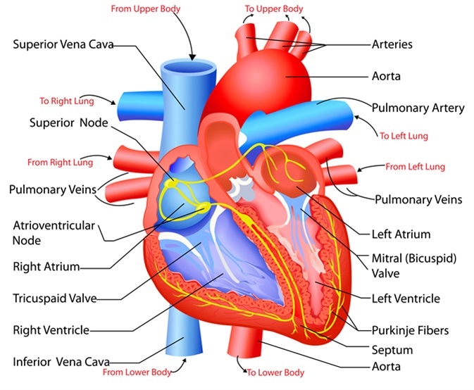
Heart structure with labels. Label the Heart Diagram | Quizlet Label the Heart Diagram | Quizlet Label the Heart 4.6 (47 reviews) + − Learn Test Match Created by bluesas9 Terms in this set (15) Superior Vena Cava ... Right Ventricle ... Left Atrium ... Atrioventricular/Tricuspid Valve ... Atrioventricular/Mitral Valve ... Septum ... Right Atrium ... Semi-lunar Valves ... Left Pulmonary Veins ... Label the HEART | Circulatory System Quiz - Quizizz 24 Questions Show answers Question 1 60 seconds Q. What is number two pointing at in the heart diagram? answer choices Right Atrium Right Ventricle Left Atrium Left Ventricle Question 2 60 seconds Q. What is number one pointing at in the heart diagram? answer choices Right Ventricle Right Pulmonary Vein Superior Franklin Inferior Vena Cava Label the heart — Science Learning Hub Label the heart Interactive Add to collection In this interactive, you can label parts of the human heart. Drag and drop the text labels onto the boxes next to the diagram. Selecting or hovering over a box will highlight each area in the diagram. pulmonary vein semilunar valve right ventricle right atrium vena cava left atrium pulmonary artery How to Draw the Internal Structure of the Heart (with Pictures) - wikiHow Make sure to label the following: Superior Vena Cava Inferior Vena Cava Pulmonary Artery Pulmonary Veins Left Ventricle Right Ventricle Left Atrium Right Atrium Mitral Valves Aortic Valves Aorta Pulmonic Valve (Optional) Tricuspid Valve (Optional) 6 To finish, label "The Human Heart" above the sketch. Tips Use pencil
Structure of the Heart | The Franklin Institute The heart consists of four chambers: two atria on the top and two ventricles on the bottom. Looking at the Valentine's Day heart, the two rounded humps at the top are rounded like the top of a lower-case "a." The bottom is shaped like a "v." Feel it working What else is inside your heart? Simple heart diagram | Simple heart diagram labeled | Human heart ... Internal structure of human heart shows four chambers viz. two atria and two ventricles and couple of blood vessels opening into them. The wall of two ventricles are strong and sturdy when compared to atria. Before we start, we shall recall the basic proportions of heart and its chambers. The right Auricle is larger than left. Heart Diagram with Labels and Detailed Explanation - Collegedunia The heart is located under the ribcage, between the lungs and above the diaphragm. It weighs about 10.5 ounces and is cone shaped in structure. It consists of the following parts: Heart Detailed Diagram Heart - Chambers There are four chambers of the heart . The upper two chambers are the auricles and the lower two are called ventricles. 8 South African Record Labels Looking To Sign New Artists 06.01.2022 · 8 Record Labels in South Africa Looking for Artistes to Sign . Here are the highest paying record labels in South Africa looking for talented new artists to sign. 1. Gallo Records. Gallo Record Company is the first South Africa record label on the list looking for new artists. Gallo Record Company has been in operation since 1926, and incorporates both Sheer Music …
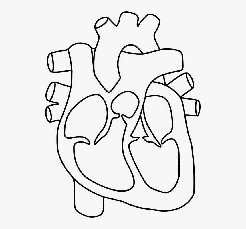
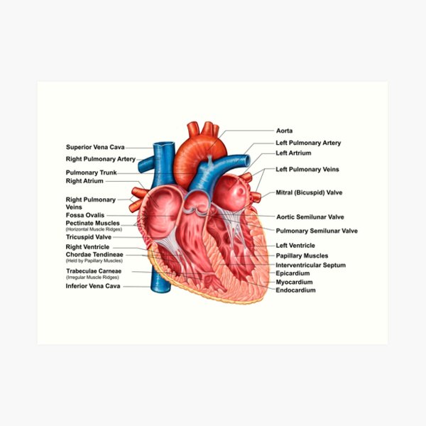
The Anatomy of the Heart, Its Structures, and Functions - ThoughtCo The heart is the organ that helps supply blood and oxygen to all parts of the body. It is divided by a partition (or septum) into two halves. The halves are, in turn, divided into four chambers. The heart is situated within the chest cavity and surrounded by a fluid-filled sac called the pericardium. This amazing muscle produces electrical ... Diagrams, quizzes and worksheets of the heart | Kenhub Worksheet showing unlabelled heart diagrams. Using our unlabeled heart diagrams, you can challenge yourself to identify the individual parts of the heart as indicated by the arrows and fill-in-the-blank spaces. This exercise will help you to identify your weak spots, so you'll know which heart structures you need to spend more time studying ... Human Heart: Label the diagram 1 worksheet Human Heart: Label the diagram 1 Study the figure carefully.Label the 10 parts of the human heart A-J. ID: 1781041 Language: ... Main content: Human Circulatory System Other contents: Human Heart Add to my workbooks (15) Download file pdf Embed in my website or blog Add to Google Classroom Add to Microsoft Teams Share through Whatsapp: Link to ... Dapagliflozin - Wikipedia Dapagliflozin, sold under the brand names Farxiga (US) and Forxiga (EU) among others, is a medication used to treat type 2 diabetes. It is also used to treat adults with certain kinds of heart failure and chronic kidney disease.. Common side effects include hypoglycaemia (low blood sugar), urinary tract infections, genital infections, and volume depletion (reduced amount of …
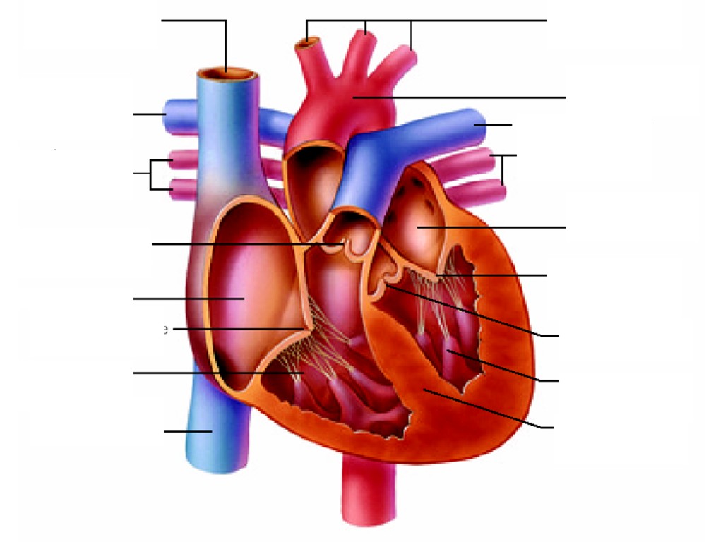
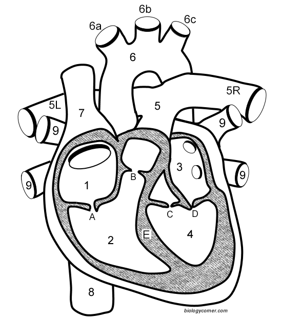
Given alongside is a diagram of the human heart showing its internal structure. Label the parts ...
Eplerenone: a blood pressure medicine used to treat heart failure It’s used to treat heart failure and reduce the risk of you having other heart problems or a stroke. It also helps to stop heart failure getting worse. It can sometimes be used to treat a condition called hyperaldosteronism. This is when your body makes too much aldosterone, a hormone that controls your blood pressure. Eplerenone comes as tablets and is only available on …

Label every structure on the figure of the heart: image ...
Labelling the heart — Science Learning Hub Labelling the heart — Science Learning Hub Labelling the heart Add to collection The heart is a muscular organ that pumps blood through the blood vessels of the circulatory system. Blood transports oxygen and nutrients to the body. It is also involved in the removal of metabolic wastes. Topics Concepts Citizen science Teacher PLD Glossary Sign in
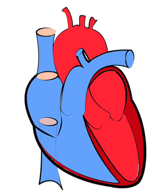
Free Heart Diagram Unlabeled, Download Free Heart Diagram ...
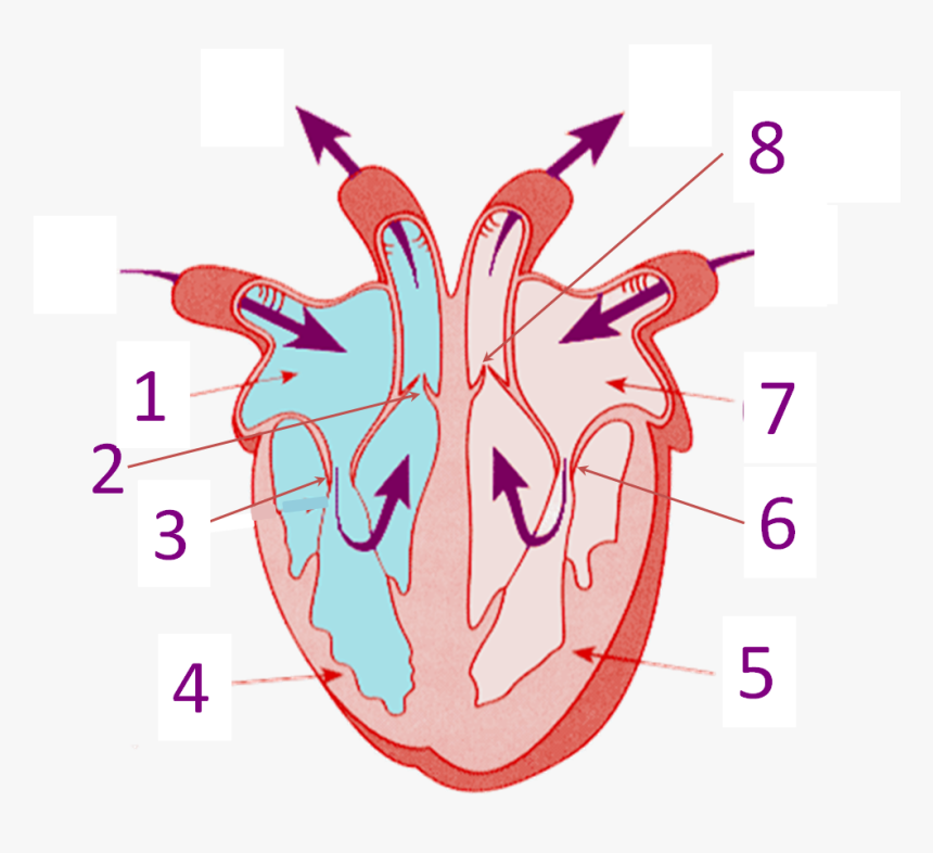
Heart Labeling Quiz: How Much You Know About Heart Labeling? Here is a Heart labeling quiz for you. The human heart is a vital organ for every human. The more healthy your heart is, the longer the chances you have of surviving, so you better take care of it. Take the following quiz to know how much you know about your heart. Questions and Answers 1. What is #1? 2. What is #2? 3. What is #3? 4. What is #4?
Conduction system of the heart: Parts and Functions | Kenhub
Structure of Heart (With Diagram) | Circulatory System | Human Physiology The heart is consisting of three layers: 1. Pericardium or outer covering layer: The heart lies in a double membranous sac of pericardium with serous fluid between the two layers. This is known as pericardial fluid. By its lubricating action, the heart can move freely or contracts and expands without any injury.
Heart Information Center: Heart Anatomy | Texas Heart Institute
Label the Heart Quiz - PurposeGames.com Ummmmmmm . . . it's pretty self explanatory . . . you label the heart. Just remember one thing - you're looking at the heart like it's in someone else so right and left are switched around. This quiz has tags. Click on the tags below to find other quizzes on the same subject. Anatomy.
13+ Heart Diagram Templates – Sample, Example, Format ...
Fellow of the American Heart Association (FAHA) For those who qualify, election as a Fellow of the American Heart Association recognizes your scientific and professional accomplishments, volunteer leadership and service. By earning the right to include the initials FAHA among your credentials, you let colleagues and patients know that you have been welcomed into one of the world’s most eminent organizations of …
Label the heart — Science Learning Hub
Human Heart (Anatomy): Diagram, Function, Chambers, Location in ... - WebMD The heart is a muscular organ about the size of a fist, located just behind and slightly left of the breastbone. The heart pumps blood through the network of arteries and veins called the...
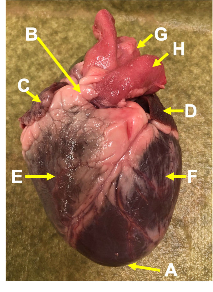
Heart Anatomy: Heart Dissection
heart structure 3D realistic interactive virtual heart structure A common misconception is that all veins carry deoxygenated blood. It is more appropriate to classify veins as vessels carrying blood to the heart. MRI anatomy of the Horse heart The three-dimensional horse and heart on the left can be rotated, zoomed, and panned by clicking and moving the mouse as indicated.
Free Heart Diagram Unlabeled, Download Free Heart Diagram ...
Human Heart - Anatomy, Functions and Facts about Heart - BYJUS To know more about the human heart structure and function, or any other related concepts such as arteries and veins, ... Practice your understanding of the heart structure. Drag and drop the correct labels to the boxes with the matching, highlighted structures. Instructions to use: Hover the mouse over one of the empty boxes. One part in the image gets highlighted. Identify the …

Structure of the heart interactive worksheet
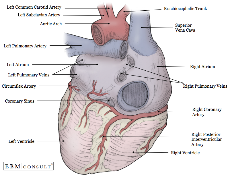
Laboratory exercise 38 heart structure figurelabels - Course Hero LABORATORY EXERCISE 38 HEART STRUCTURE Figure Labels FIG. 38.1 1. Aorta 7. Pulmonary trunk (artery) 2. Superior vena cava 8. Left atrium 3. Right atrium 9. Left coronary artery 4. Right coronary artery 10. Great cardiac vein 5. Right ventricle 11. Left ventricle 6. Inferior vena cava FIG. 38.2 1. Aorta 6. Apex 2. Left pulmonary artery 7.
Heart Structure Without Label, HD Png Download - kindpng
Simple heart diagram | Simple heart diagram labeled | Human ... - Pinterest Simple heart diagram labeled with accurate labels. Most frequent question in exam to draw human heart diagram with labels. You can learn diagram of heart with labels and easy simple heart anatomy with heart structure. Learn to draw Simple heart diagram in very simple way and also learn simple heart anatomy. Pharmacy Images 386 followers
Learn the Anatomy of the Heart
The Heart vs. The Mind (scientific explanation) - Cognition Today 16.02.2018 · Categorizing 2 different things with labels like the heart & mind also gives them unique meaning, just because of the categorization with labels. That means, we tend to give this classification additional meaning. People generally care just about that additional meaning. That is, the label represents more than the thing that is labeled. For example, look at the concept of …
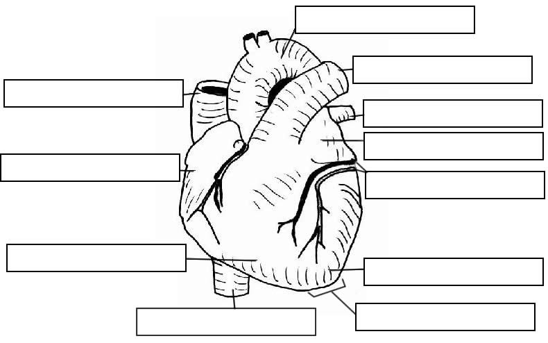
Anatomy: Heart (External)
Structure and Function of the Heart - News-Medical.net Structure of the heart The heart wall is composed of three layers, including the outer epicardium (thin layer), middle myocardium (thick layer), and innermost endocardium (thin layer). The...
Anatomy of heart interior with labels — semilunar valve ...
Carbohydrates | American Heart Association 16.04.2018 · Carbohydrates are either called simple or complex, depending on the food’s chemical structure and how quickly the sugar is digested and absorbed. The type of carbohydrates that you eat makes a difference – Foods that contain high amounts of simple sugars, especially fructose raise triglyceride levels. Triglycerides (or blood fats) are an …
Label the heart - Teaching resources
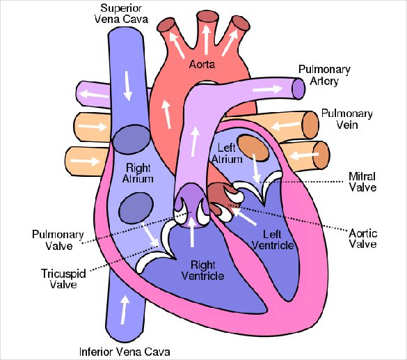
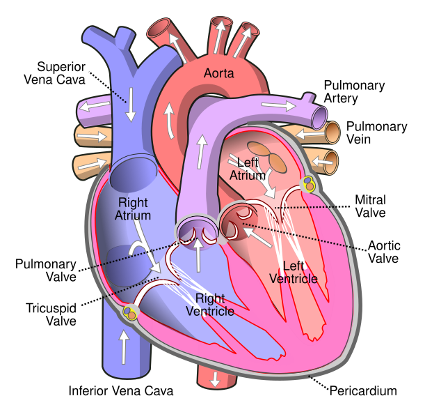
Heart Diagram for Kids - Bodytomy As you can see in the diagram of the heart, that heart is divided in four chambers, namely, right atrium, left atrium, right ventricle and left ventricle. Each of the chambers is separated by a muscle wall known as Septum. The left side of the heart receives oxygen rich blood from the lungs and pumps it out the whole body.

Heart Lab Flashcards | Quizlet
Heart: Anatomy and Function - Cleveland Clinic Heart. Your heart is the main organ of your cardiovascular system, a network of blood vessels that pumps blood throughout your body. It also works with other body systems to control your heart rate and blood pressure. Your family history, personal health history and lifestyle all affect how well your heart works. Appointments 800.659.7822.
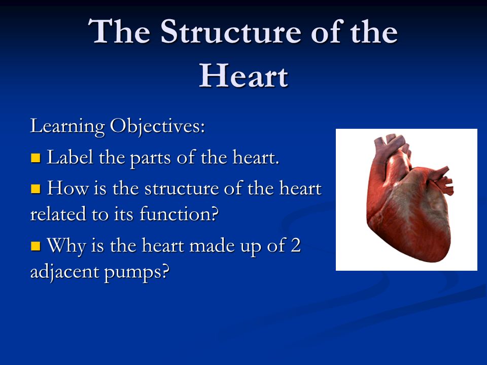
The Structure of the Heart Learning Objectives: Label the ...
PDF Heart Structure - Indiana The heart is an organ about the size of a fist. It is made of muscle and pumps blood through the body. Tube-like structures called blood vessels carry blood through the body and heart. The heart and blood vessels make up the cardiovascular system. Structure of the Heart The heart has four chambers: two upper chambers call
Heart Diagram Answer Key.indd
Heart anatomy: Structure, valves, coronary vessels | Kenhub Heart anatomy. The heart has five surfaces: base (posterior), diaphragmatic (inferior), sternocostal (anterior), and left and right pulmonary surfaces. It also has several margins: right, left, superior, and inferior: The right margin is the small section of the right atrium that extends between the superior and inferior vena cava .
Heart Anatomy Review
Lab 44- Heart Structure Flashcards | Quizlet Right side: 1.)Ligamentrum. 2.)left pulmonary artery. 3.)Pulmonary trunk. 4.)Left pulmonary veins. 5.)Auricle of left atrium. 6.)Grat cardiac vein. 7.)Anterior interventricular artery. Label the posterior heart structures by clicking and dragging the labels to the correct location.
The Human Heart
RiSA planning overhaul of Samas 'structure, system and … 01.09.2022 · Sibisi attributed this to "less than optimal" engagement with their members (record companies and artist-led labels) because of Covid-19 and the government-imposed restrictions. "The Samas rely on a system in which our members' review, amend and confirm the rules, categories, and judges. This system is reviewed annually by our members and ...
Heart Anatomy: Labeled Diagram, Structures, Blood Flow ...
Structure of the Heart | SEER Training - National Cancer Institute Layers of the Heart Wall Three layers of tissue form the heart wall. The outer layer of the heart wall is the epicardium, the middle layer is the myocardium, and the inner layer is the endocardium. Chambers of the Heart The internal cavity of the heart is divided into four chambers: Right atrium Right ventricle Left atrium Left ventricle

Pin on Anatomy
Heart Diagram with Labels and Detailed Explanation - BYJUS Diagram of Heart. The human heart is the most crucial organ of the human body. It pumps blood from the heart to different parts of the body and back to the heart. The most common heart attack symptoms or warning signs are chest pain, breathlessness, nausea, sweating etc. The diagram of heart is beneficial for Class 10 and 12 and is frequently ...

Simple heart diagram | Simple heart diagram labeled | Human ...
Anatomy, Health, Heart, Human, Science - Human Heart Diagram ...
Heart Diagram with Labels and Detailed Explanation
Structure and Function of the Heart
Anatomy of heart interior, frontal section. Art Print by StocktrekImages

Pin on MCAT
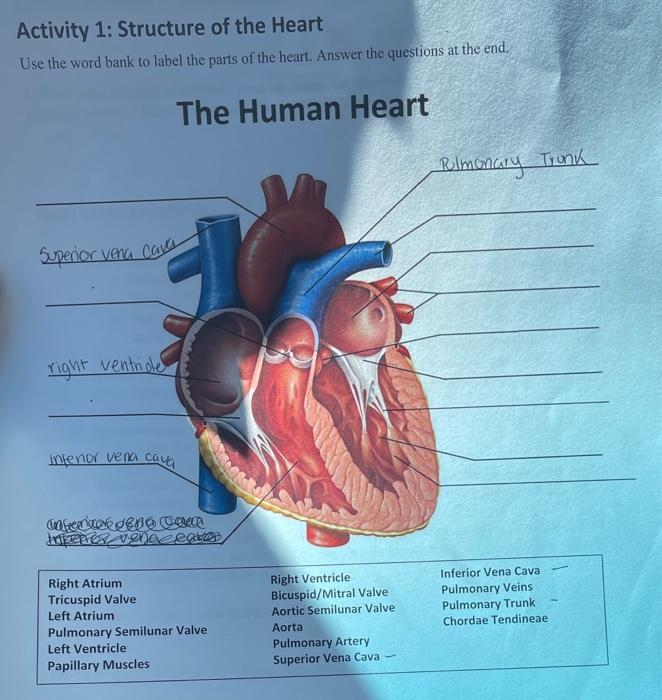
Solved Activity 1: Structure of the Heart Use the word bank ...

4,091 Human Heart Diagram Stock Photos, Pictures & Royalty ...

Heart Anatomy | Anatomy and Physiology II
Heart Anatomy: Labeled Diagram, Structures, Blood Flow ...
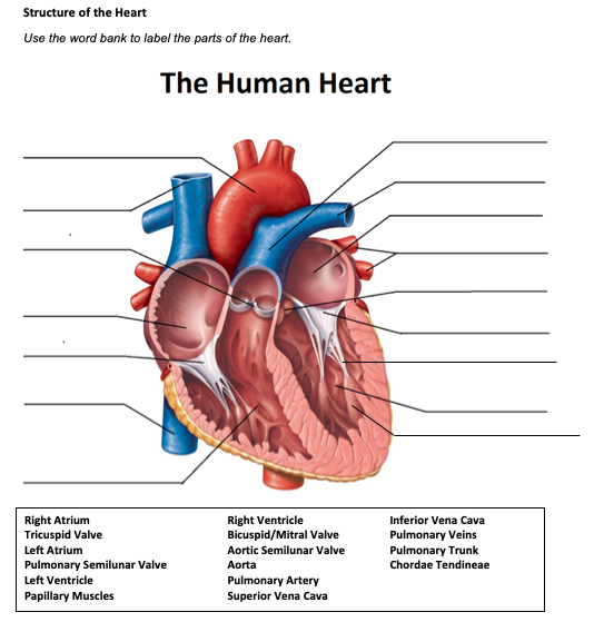
Solved Structure of the Heart Use the word bank to label the ...
Notes: Heart and Circulatory System

Sketch Of Human Heart Anatomy With Hand Written Labels Stock ...
Congenital Heart Defects - How the Heart Works | CDC
File:Diagram of the human heart (cropped).svg - Wikimedia Commons

4,091 Human Heart Diagram Stock Photos, Pictures & Royalty ...

Pin on Paramedic Study Guide
The heart: Anatomy, how it works, and more
Post a Comment for "40 heart structure with labels"